Do you need advice? +420 545 235 668
")
")


Posterior Abdominal wall
Catalog number: EM270-MP1300
Product availability: Delivery date HERE
Weight:
6 Kg
Description
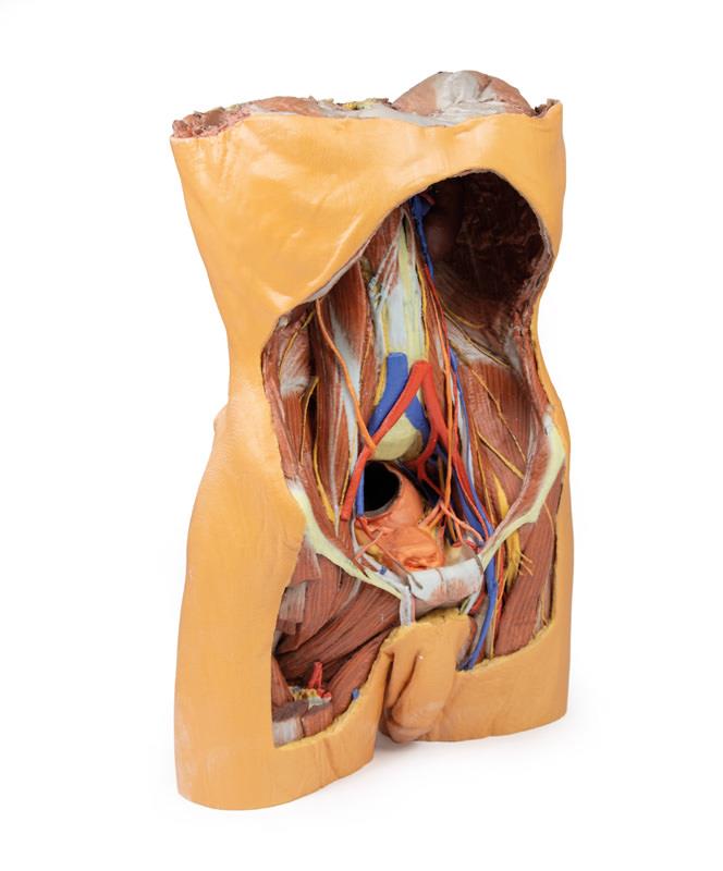
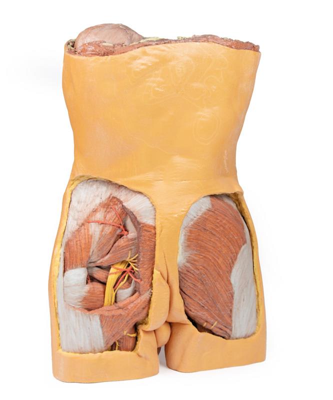
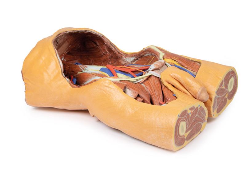
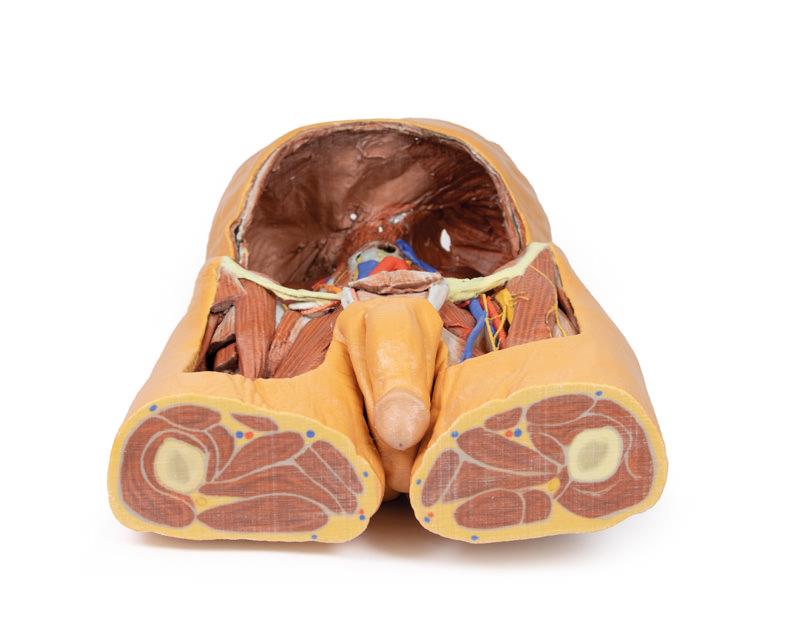
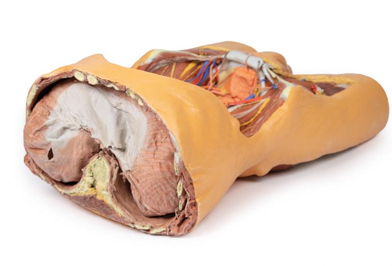
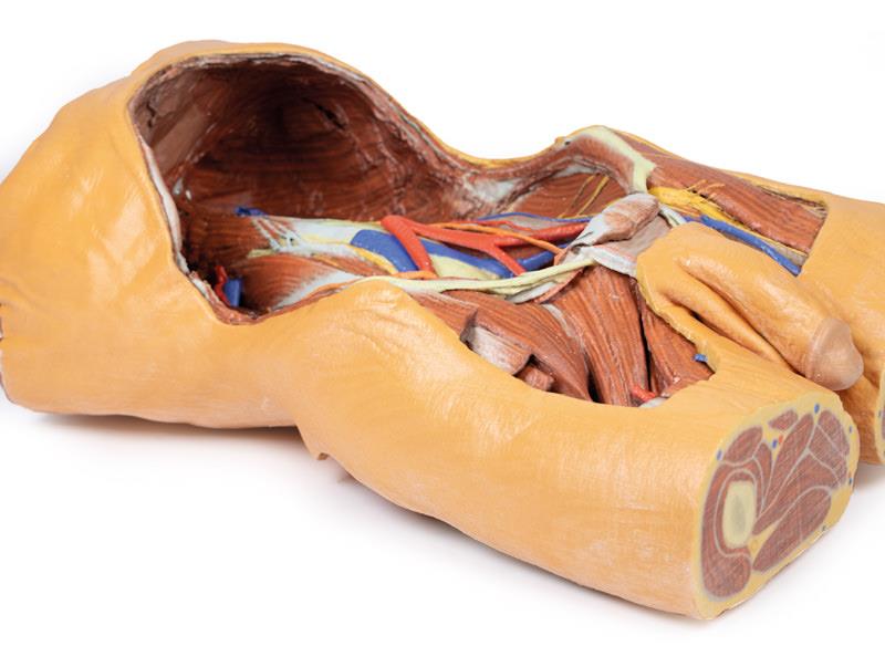
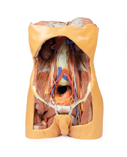
A large, multipart 3D printed specimen offering a detailed anatomical view of the the entire male posterior abdominal wall from the diaphragm to the pelvic brim. Ideal for study of the anatomy of the pelvic area and proximal thigh.
The 3D Human Anatomy Series is the result of a collaboration between Monash University in Australia and German manufacturer Erler Zimmer. The 3D printed human anatomy models in this collection are highly accurate colour-augmented representations, designed to substitute the use of real human cadavers in anatomy education. Each model has been developed from radiographic data or cadaver specimins, providing a highly cost effective and ethical solution to meet the needs of medical, allied health and biological science educators.
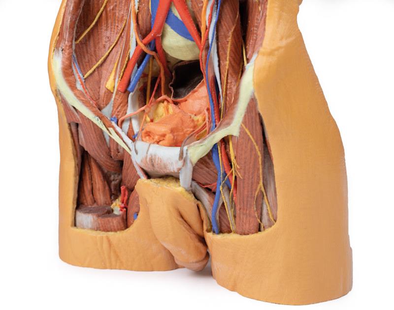
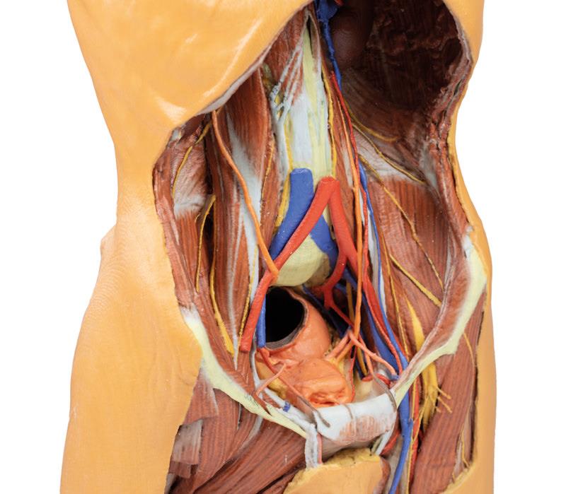
In this posterior abdominal wall specimim, the parietal peritoneum has been removed from the posterior abdominal wall to expose the muscular wall including the psoas, the quadratus lumborum, transversus abdominus, and the iliacus below the iliac crest. The muscular portions of the dome shaped diaphragm are clearly distinct from the central tendon. The fibres originate from the circumference of the internal walls of the bony thorax at its margin (sternal fibres, costal portion, lumbar portion). The origins of the diaphragm and the left and right crura are clearly identifiable originating from the vertebral bodies (L1-L3 on the right and L1-L2 on the left. The crura are connected by a tendinous band, the median arcuate ligament, which arches in front of the aorta; however in this specimen the aorta has been removed. The fibres of the diaphragm arising from the tendinous arches over psoas and the lateral arcuate ligaments are partly hidden by the kidneys. The oesophageal opening through the arching fibres of the right crus is present above (level of T10) and to the left of the aortic opening (level of T12). The opening in the central tendon that transmits the inferior vena cava (level of T8/9 intervertebral disc).
The somatic nerves of the posterior abdominal wall are clearly identifiable and consist of from above downwards – the subcostal, the iliohypogastric and ilioinguinal nerves lie on the quadratus lumborum (in this individual they arise together and– this can often occur and they split later in abdominal muscle layers), the lateral cutaneous nerve of thigh, the femoral lying in the groove between psoas and iliacus), and the genitofemoral nerve lying superficially upon psoas. The sympathetic trunks can be seen descending lateral to the lumbar vertebral bodies.
The aorta and inferior cava are transected around the level of L3 vertebral body. The aortic bifurcation into the right and left common iliac arteries is slightly higher than normal.
Finally, the kidneys have dissected from the peri- and pararenal fat of the posterior abdominal wall. The renal vessels (arteries anteriorly, veins posteriorly) have been preserved but as the aorta and inferior cava have been removed this does display the origin and arrangements of these vessels fully. The more inferior position of the right kidney is clearly visible and the ureters can be seen emerging from the hilum and descending initially lateral to psoas, then anterior to this muscle before crossing pelvic brim anterior to the bifurcation of the common iliac arteries to reach the true pelvis.