Do you need advice? +420 545 235 668
")
")


Female Pelvis with Ligaments, Vessels, Nerves, Pelvic Floor, Organs - 3B Smart Anatomy
Catalog number: AM110-0580
Product availability: Delivery date HERE
Weight:
3 Kg
Price incl. VAT18 694,50 CZK
Description
Your advantages with all 3B Smart Anatomy models:
Free warranty extension from 3 to 5 years
Free access to 3B Smart Anatomy courses in the award-winning Complete Anatomy
Includes 11 3B Smart Anatomy courses with 23 lectures and 117 different views of interactive virtual models. Also includes 39 quizzes
To unlock these benefits, simply scan the label and register your 3B Smart Anatomy model online. All 3B Smart Anatomy features are completely free of charge for you. Read more about the concept of "Virtual meets Reality" developed by 3B Scientific for you to make learning human anatomy even more effective across all media.
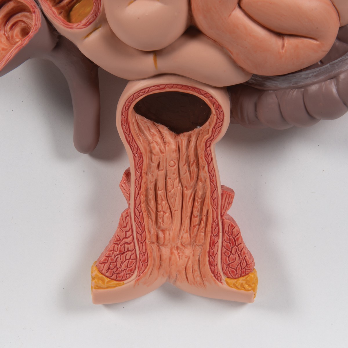
This life size six part model of a female pelvis represents detailed information about the topography of bones, ligaments, vessels, nerves, pelvic floor muscles and female genital organs. It presents the whole pelvic floor with partially removable midsagitally sectioned external anal sphincter, external urethral sphincter, deep and superficial transverse perineal and bulbospongiosus.
The rectum, uterus with fallopian tubes, ovaries and vagina are also removable and can be disassembled into both halves by midsagital section. The right pelvic half demonstrates the divisions and topographical anatomy of the common iliac artery, the external and internal artery and also of the common iliac vein and the external iliac vein. The right sacral plexus, right sciatic nerve and right pudendal nerve are also shown.
Bones and ligaments presented: Two hip bones, the pubic symphysis, the sacrum and the coccyx, the fifth lumbar vertebra with intervertebral disc. A midsagital section through the fifth lumbar vertebra, sacrum and coccyx, allow both halves of the pelvis to be disassembled revealing a part of the cauda equina in the vertebral canal. The left half of the fifth lumbar vertebral body is removable. The right half of the model shows the following pelvic ligaments: inguinal ligament, sacrotuberous ligament, sacrospinous ligament, anterior sacroiliac ligaments, iliolumbar ligament, anterior longitudinal ligament, interosseous sacroiliac ligament, posterior sacroiliac ligament and obturator.
This model is great for detailed study of the female genital and pelvic anatomy.
Free warranty extension from 3 to 5 years
Free access to 3B Smart Anatomy courses in the award-winning Complete Anatomy
Includes 11 3B Smart Anatomy courses with 23 lectures and 117 different views of interactive virtual models. Also includes 39 quizzes
To unlock these benefits, simply scan the label and register your 3B Smart Anatomy model online. All 3B Smart Anatomy features are completely free of charge for you. Read more about the concept of "Virtual meets Reality" developed by 3B Scientific for you to make learning human anatomy even more effective across all media.
This life size six part model of a female pelvis represents detailed information about the topography of bones, ligaments, vessels, nerves, pelvic floor muscles and female genital organs. It presents the whole pelvic floor with partially removable midsagitally sectioned external anal sphincter, external urethral sphincter, deep and superficial transverse perineal and bulbospongiosus.
The rectum, uterus with fallopian tubes, ovaries and vagina are also removable and can be disassembled into both halves by midsagital section. The right pelvic half demonstrates the divisions and topographical anatomy of the common iliac artery, the external and internal artery and also of the common iliac vein and the external iliac vein. The right sacral plexus, right sciatic nerve and right pudendal nerve are also shown.
Bones and ligaments presented: Two hip bones, the pubic symphysis, the sacrum and the coccyx, the fifth lumbar vertebra with intervertebral disc. A midsagital section through the fifth lumbar vertebra, sacrum and coccyx, allow both halves of the pelvis to be disassembled revealing a part of the cauda equina in the vertebral canal. The left half of the fifth lumbar vertebral body is removable. The right half of the model shows the following pelvic ligaments: inguinal ligament, sacrotuberous ligament, sacrospinous ligament, anterior sacroiliac ligaments, iliolumbar ligament, anterior longitudinal ligament, interosseous sacroiliac ligament, posterior sacroiliac ligament and obturator.
This model is great for detailed study of the female genital and pelvic anatomy.