Do you need advice? +420 545 235 668
")
")


Heart internal structures
Catalog number: EM270-MP1715
Product availability: Delivery date HERE
Weight:
1 Kg
Description
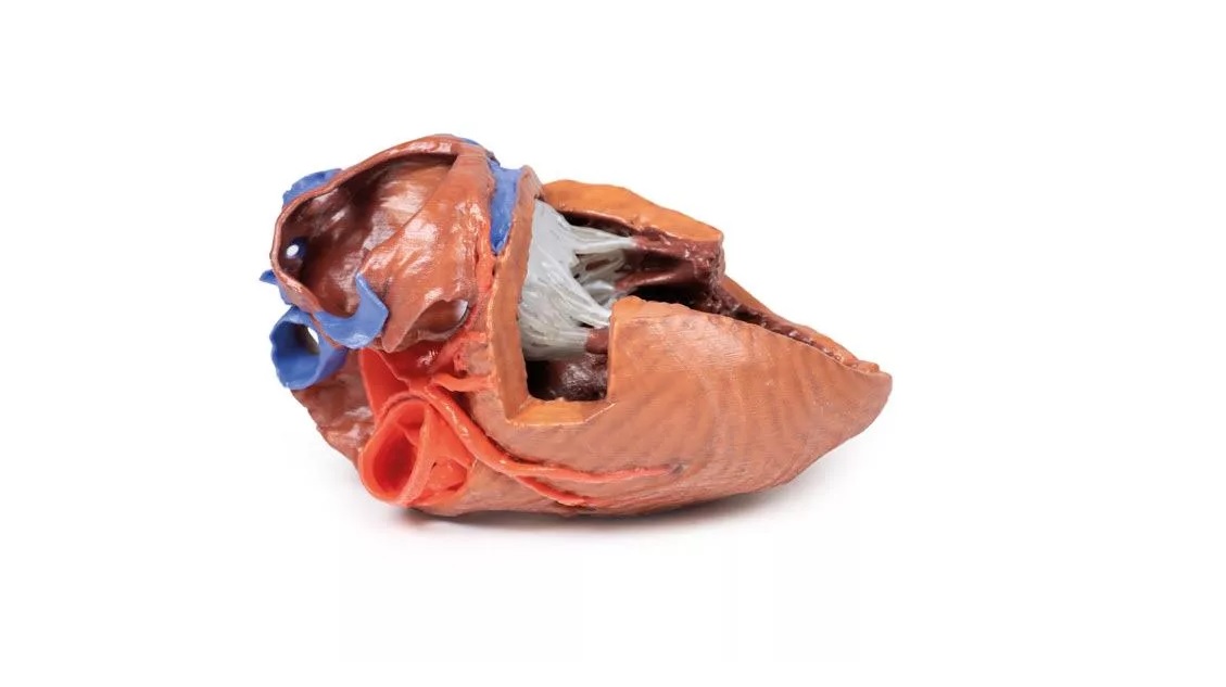
This 3D printed heart has been dissected to display the internal structures of the chambers. At the base of the heart the termination of the superior vena cava is preserved entering the right atrium. Part of the inferior vena cava is also preserved on the inferior aspect of the right atrium; however, most of the vessel lumen and much of the anterior wall has been removed to expose the pectinate muscles of the right auricle and the fossa ovalis (which is nearly translucent in the 3D print). The anterior wall of the right ventricle has also been removed to expose the right atrioventricular valve and its three cusps (anterior, posterior, and septal), including the chordae tendineae connecting them to respective papillary muscles projecting
from trabeculae carneae (including a septomarginal trabecula entering the anterior papillary muscle from the interventricular septum).
from trabeculae carneae (including a septomarginal trabecula entering the anterior papillary muscle from the interventricular septum).